

Yes — ultrasound and TVS are safe during pregnancy. Decades of clinical use and major bodies including ACOG and AIUM confirm there is no evidence that properly performed ultrasound causes miscarriage. Ultrasound uses sound waves, not radiation. TVS does not touch the baby or placenta. When miscarriage seems to “follow” a scan, the scan is detecting a problem that was already present — not causing it.
📋 What This Guide Covers
- What ultrasound & TVS actually are
- Can ultrasound or TVS cause miscarriage?
- Why “miscarriage after scan” is a false association
- Types of pregnancy ultrasound — compared
- How many scans are recommended (and when)
- The ALARA principle — what safe really means
- The honest Doppler caveat
- 3D/4D and keepsake scans
- If you’re an IVF patient — what’s normal
- When to call your doctor after a scan
- A note from Dr. Shradha, Patna
- FAQs
What Ultrasound & TVS Actually Are
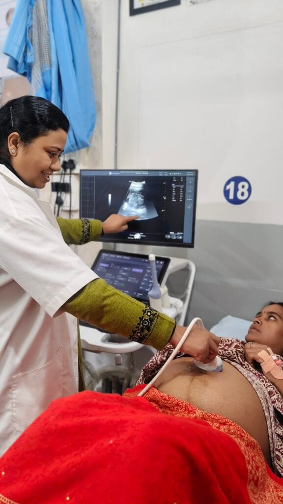
Ultrasound (sonography) is a medical imaging test that uses sound waves — not radiation, not X-rays, not electricity — to create a picture of the baby and the womb. A small device called a transducer sends inaudible high-frequency sound waves into the body; the echoes that bounce back are translated into the live image you see on the screen.
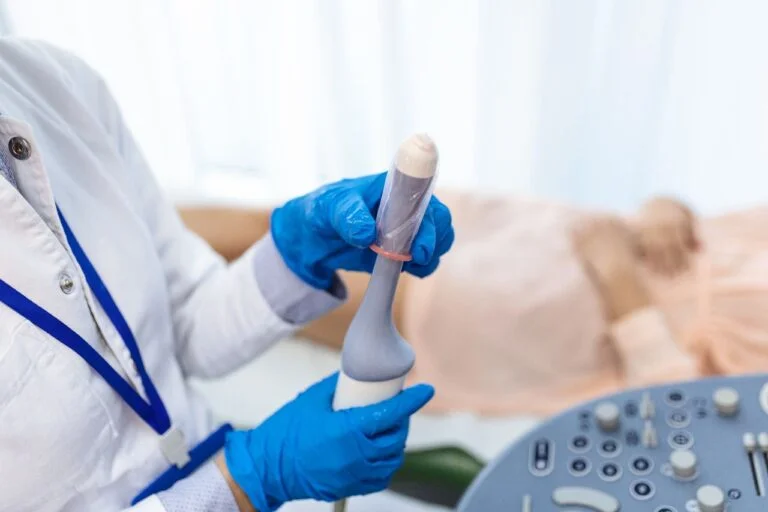
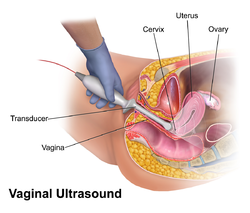
TVS (Transvaginal Sonography) is a specific type of ultrasound used in early pregnancy. A thin, smooth, gel-covered probe is gently placed in the vagina to get a closer view of the uterus and ovaries. The probe stops in the vagina; it does not enter the cervix, the uterus, or anywhere near the baby. It does not touch the placenta or the embryo. It is essentially an internal angle on the same safe sound-wave technology.
Neither test involves needles, medication, radiation, or electrical current. Both are non-invasive imaging — among the gentlest tests in all of medicine.
Can Ultrasound or TVS Cause Miscarriage?
The clear, honest answer is no. There is no published medical evidence — across decades of routine use and many large studies — that properly performed ultrasound or TVS causes miscarriage. Major authoritative bodies that have reviewed the evidence in detail agree:
- ACOG (American College of Obstetricians and Gynecologists) calls ultrasound the preferred modality for verifying early pregnancy and considers it safe throughout pregnancy when performed for a medical reason.
- AIUM (American Institute of Ultrasound in Medicine) states that “no biological effects have been independently confirmed in patients exposed to typical present-day diagnostic ultrasound” — while reinforcing the prudent ALARA principle (more on that below).
- NHS (UK) describes ultrasound as safe for both mother and baby when performed by trained personnel.
In simple language: there is no mechanism by which a properly used ultrasound can cause a pregnancy to end. The energy levels used are far below anything that could harm tissue, the probe never touches the baby, and the evidence base spanning millions of pregnancies is consistent.
Why “Miscarriage After Scan” Is a False Association
This is the most important paragraph in this article — please read it slowly. Most early miscarriages (about 80% of all miscarriages) happen before 12 weeks of pregnancy. The first ultrasound scan in most pregnancies is also done in this same window — between 6 and 12 weeks. So chronologically, the scan and a miscarriage often occur close together.
But “close together” is not the same as “caused by.” This is what statisticians call a false association — two things happening near each other in time, where one did not cause the other. In almost every case where a miscarriage is discovered around the time of a scan, the scan is the moment the problem was detected, not the moment it was caused. The pregnancy was already not progressing — the scan simply revealed it. Without the scan, the loss would have shown itself a few days or weeks later as bleeding or cramping.
This is why we sometimes describe ultrasound as the messenger of bad news, not the cause of it. And it’s why declining a scan out of fear can be more harmful than helpful — silent problems like ectopic pregnancy or missed miscarriage can go undetected without imaging, and these are situations where early recognition genuinely saves lives.
What are the Different Types of Pregnancy Ultrasound A Doctor Recommends?
| Type | When Used | What It Checks | Safe? |
|---|---|---|---|
| TVS (transvaginal) | First trimester | Pregnancy location, viability, heartbeat, ovary check | ✅ Yes |
| Abdominal (B-mode) | All trimesters, mainly 2nd & 3rd | Baby’s size, position, anatomy, placenta, fluid | ✅ Yes |
| Doppler (colour/power) | Mainly 2nd–3rd trimester | Blood flow in cord, placenta, fetal vessels | ✅ Yes when medically indicated |
| Spectral Doppler (“hear the heartbeat”) | When clinically needed | Higher acoustic output; use cautiously in 1st trimester (see AIUM caveat below) | ⚠️ Yes, with caution in 1st trimester |
| 3D / 4D scans | 2nd / 3rd trimester | Detailed surface images, facial features | ✅ Safe medically; not for keepsakes (see below) |
How Many Scans Are Recommended (and When) by the Doctor and Why?
A common worry: “Am I having too many scans?” Here’s the honest breakdown of when scans are typically done in a low-risk pregnancy. Your obstetrician will personalise this for you.
| When | Scan | Why It’s Done |
|---|---|---|
| 6–8 weeks | TVS — early viability scan | Confirm pregnancy is in the uterus (not ectopic), confirm heartbeat, and accurate dating |
| 11–14 weeks | NT (nuchal translucency) scan | First-trimester screening assesses the risk of chromosomal conditions |
| 18–22 weeks | Anomaly scan (TIFFA / fetal anatomy) | Detailed check of the baby’s anatomy, organs, growth |
| 28–32 weeks | Growth scan | Baby’s growth, placenta position, and fluid levels |
| 36+ weeks (if needed) | Term/well-being scan | Position, fluid, Doppler if indicated |
Most uncomplicated pregnancies have 3–5 scans total across nine months. Additional scans are added when there’s a clinical reason — bleeding, pain, suspected growth issues, or a high-risk pregnancy (including IVF pregnancies in the first trimester, which we’ll cover separately). Each extra scan is for a reason — never for “extra reassurance” with no purpose.
The ALARA Principle — What “Safe” Really Means
Doctors and sonographers worldwide follow a principle called ALARA — As Low As Reasonably Achievable. It simply means: even though ultrasound has no known harm at diagnostic levels, we still use the shortest exposure time and the lowest power setting needed to get the clinical information we need. Nothing more.
In simple words, we don’t do scans for no reason, we don’t linger longer than needed, and we set the equipment as gently as possible. ALARA is not because ultrasound is dangerous — it’s because good medicine is always conservative with energy exposure, even when the energy in question has no proven harm. This is why you’ll sometimes hear us discourage non-medical scans (see 3D/4D below) — they don’t add medical value, but they do add exposure.
The Honest Doppler Caveat
This is the one nuance most blog posts skip — but you deserve to know it, because honest writing builds trust. Spectral Doppler ultrasound (used to “hear” the heartbeat as an audio sound, or to measure exact blood-flow velocity) uses higher acoustic output than standard B-mode imaging. AIUM specifically advises that spectral Doppler in the first trimester should be used cautiously — only when there’s a clear clinical reason, with short duration and low power settings.
This does not mean Doppler is harmful — there is no evidence of harm in clinical practice. It simply means it’s a tool we use thoughtfully, not casually. Standard B-mode TVS to confirm a heartbeat visually (which is what’s done routinely) is entirely safe. The audio “thump-thump” sound, when used, is brief. If your sonographer follows AIUM/ACOG guidance — and ours does — there’s nothing to worry about. Being honest about the caveat is precisely what makes the overall reassurance trustworthy.
3D / 4D and “Keepsake” Scans
Many couples are tempted by 3D and 4D scans — the beautiful images of the baby’s face, sometimes offered at non-medical centres or shopping-mall studios. Medical 3D/4D scans, ordered by your doctor for a clinical reason, are safe.
However, both AIUM and the FDA advise against non-medical “keepsake” ultrasound scans done purely for entertainment or souvenir photographs. The reason is not that they’re dangerous — it’s that there’s no medical benefit to justify the extra exposure, the operators may not be medically trained, and the scans can last much longer than clinically necessary. ALARA again. If you want a 3D image, ask your doctor to capture one during your routine anomaly scan — that’s the safer way to have your special photograph.
If You’re an IVF Patient — What’s Normal
This deserves a section of its own. IVF patients typically have more first-trimester scans than naturally conceived pregnancies — often 4–6 TVS scans before 12 weeks. This can sound alarming if you’ve just read the rest of this article. It isn’t. Here’s why:
- Early scans confirm the pregnancy is in the uterus (not ectopic) — particularly important after embryo transfer.
- Heartbeat is checked at the right developmental milestones (typically around week 6 and again around week 8).
- For IVF patients carrying twins or higher-order pregnancies, both sacs need monitoring.
- Progesterone support is being managed, and early reassurance scans guide dosing decisions.
Every single one of these scans is medically indicated. They are not “extra” — they are exactly the right level of monitoring for an IVF-conceived pregnancy. If you’d like to understand your due date and milestones, the Due Date Calculator shows when each scan typically falls. The frequency is normal, the safety is the same, and the reassurance these scans offer is part of how IVF pregnancies become healthy, term pregnancies.
When to Call Your Doctor After a Scan
- Heavy vaginal bleeding (soaking a pad in an hour or less)
- Severe, persistent abdominal or pelvic pain
- Fainting, dizziness, or feeling extremely unwell
- Fever above 38°C / 100.4°F
- Passing tissue or clots
A small amount of pink or brown spotting in the 24 hours after a TVS is occasionally normal — the cervix is sensitive in pregnancy and can spot from gentle contact. It is not caused by the scan harming the pregnancy. Anything heavier than light spotting, however, warrants a call. We always prefer being called over not being called.
A Note from Dr. Shradha, Patna
At Shradha IVF & Maternity, every pregnancy under our care — whether IVF or natural — is personally overseen by Dr Shradha, with ultrasound performed by trained sonographers in accordance with ACOG and AIUM guidance. If anything in this article raises a question for you, please ask us directly. Reassurance is part of care, and you deserve a clear, honest answer.
FAQs Related to Ultrasounds
No. There is no medical evidence that properly performed ultrasound causes miscarriage. ACOG, AIUM, and NHS all confirm ultrasound is safe in pregnancy. When miscarriage seems to follow a scan, the scan is detecting a problem that was already present — not causing it.
No. TVS is safe in pregnancy. The probe stays in the vagina and does not enter the cervix, uterus, or touch the baby. Decades of clinical use show no link between TVS and miscarriage. It is often the safest way to confirm a healthy early pregnancy.
Far far away, behind the word mountains, far from the countries Vokalia and Consonantia, there live the blind texts. Separated they live in Bookmarksgrove right at the coast
Yes. TVS is the preferred imaging method in early pregnancy because it gives clearer images than abdominal ultrasound at this stage. It is gentle, brief, and uses the same harmless sound waves. ACOG considers it the standard for confirming early pregnancy viability.
Most low-risk pregnancies have 3–5 scans across the nine months. IVF pregnancies may have more in the first trimester. There is no fixed "maximum safe number" — extra scans are added only when medically indicated. Each scan follows the ALARA principle to keep exposure minimal.
Yes, a small amount of pink or brown spotting in the 24 hours after a TVS can happen because the cervix is sensitive in pregnancy. This is not caused by harm to the pregnancy. Heavy bleeding, severe pain, or passing clots warrants an immediate call to your doctor.
No, please don't. Skipping a recommended scan is riskier than having it. Silent problems like ectopic pregnancy or missed miscarriage can be life-threatening if undetected. Talk to your doctor about your worry — but don't decide based on fear or rumours alone.
Don’t Let Fear Decide Your Care
If a recent scan, a relative’s story, or something on WhatsApp has made you anxious — please don’t carry that worry alone. A short, honest conversation with Dr. Shradha will give you a clearer answer than any forum or video. The first consultation is free.
Book a Free Consultation → 💬 WhatsApp Us